Introduction: More Than Just Hearing
When we think of the ear, we immediately think of music, conversations, and the bustling noise of the world around us. However, the human ear is actually a dual-purpose biological marvel. It is a highly complex sensory organ responsible not only for hearing, but also for maintaining our entire sense of balance and spatial orientation.
To achieve this, the ear relies on an incredible sequence of physics and neurology: capturing mechanical sound waves from the air, physically amplifying them, transferring them into a fluid medium, and finally converting them into electrical nerve impulses that the brain can decode.
Let us open the archives and follow the journey of sound through the Outer, Middle, and Inner Ear.
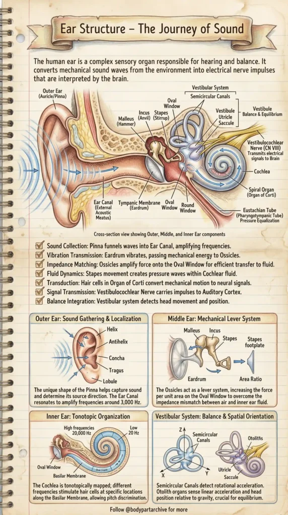
Phase 1: The Outer Ear (Sound Gathering)
The journey begins on the outside of your head. The visible part of the ear, known as the Auricle or Pinna, acts as a biological acoustic funnel.
If you look closely at the “Sound Gathering & Localization” panel, you’ll see the pinna is not just a random flap of skin. Its unique ridges and folds—including the Helix, Antihelix, Concha, and Tragus—are precisely shaped to capture sound waves and determine their directional source.
Once captured, the sound is funneled down the Ear Canal (External Acoustic Meatus). This canal is perfectly tuned to resonate and naturally amplify certain crucial frequencies (around 3,000 Hz, which is the exact range of human speech) before the sound crashes into the eardrum.
Phase 2: The Middle Ear (Mechanical Amplification)
At the end of the ear canal sits the Tympanic Membrane (Eardrum). When sound waves hit this tight membrane, it begins to vibrate, passing the mechanical energy into the Middle Ear.
Here we find the smallest bones in the human body, the Ossicles:
- Malleus (Hammer)
- Incus (Anvil)
- Stapes (Stirrup)
Why do we need these bones? The inner ear, where the actual hearing sensors live, is filled with fluid. Sound waves travel easily through the air but bounce right off fluid (a physics concept called impedance mismatch). To force the sound into the fluid, the body uses the ossicles as a Mechanical Lever System. By taking the large vibrations of the eardrum and concentrating them onto the tiny footplate of the Stapes, the middle ear drastically amplifies the force, allowing the sound to punch through the Oval Window into the inner ear.
Clinical Note: The middle ear is also home to the Eustachian Tube, which connects down to the throat. Its job is pressure equalization—which is why your ears “pop” on airplanes when you swallow.
Phase 3: The Inner Ear – Hearing (The Cochlea)
Once the Stapes pushes on the Oval Window, it creates fluid dynamics—literal waves of pressure traveling through the fluid of the Inner Ear.
This brings us to the Cochlea, a snail-shell-like structure. Inside the cochlea is the Organ of Corti, which contains microscopic hair cells. As the fluid waves wash over these hair cells, they bend. This mechanical motion triggers the “Transduction” process, converting the wave into electrical neural signals.
What is truly mind-blowing is how the cochlea decodes pitch, known as Tonotopic Organization. The membrane inside the cochlea is mapped out like a piano. High-frequency sounds (up to 20,000 Hz) stimulate the stiff hair cells right at the base near the Oval Window. Low-frequency bass sounds travel all the way to the flexible apex before triggering a signal. This is how your brain instantly distinguishes a screech from a rumble.
Phase 4: The Inner Ear – Balance (The Vestibular System)
Attached directly to the cochlea is the Vestibular System, your body’s biological gyroscope. This system has nothing to do with hearing and everything to do with spatial orientation.
It consists of two main parts:
The Semicircular Canals: Three fluid-filled loops oriented in the X, Y, and Z axes. When you turn or spin your head, the fluid inside sloshes around, allowing your brain to detect “rotational acceleration”.
The Otolith Organs (Utricle & Saccule): Located below the canals, these sac-like structures contain tiny, heavy calcium crystals called otoliths. Because they are heavy, gravity constantly pulls on them, allowing your body to sense “linear acceleration” and the exact position of your head relative to gravity.
Finally, all this massive amount of data—both the electrical sound signals from the Cochlea and the balance signals from the Vestibular system—are bundled together into the Vestibulocochlear Nerve (Cranial Nerve VIII) and sent directly into your brain for processing.
Conclusion
The ear is an absolute masterclass in biomechanical engineering. From the acoustic funnel of the pinna and the lever system of the ossicles to the 3D gyroscope of the vestibular system, it perfectly bridges our internal consciousness with the external physical world. Protecting your hearing from extremely loud noises doesn’t just save your “ears”—it preserves one of the most sophisticated neurological instruments in nature.