Disclosure: As an Amazon Associate, Body Part Archive earns from qualifying purchases. This means if you click on a link and buy something, we may earn a small commission at no extra cost to you.
Introduction: The Ultimate Multi-Tasker
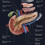
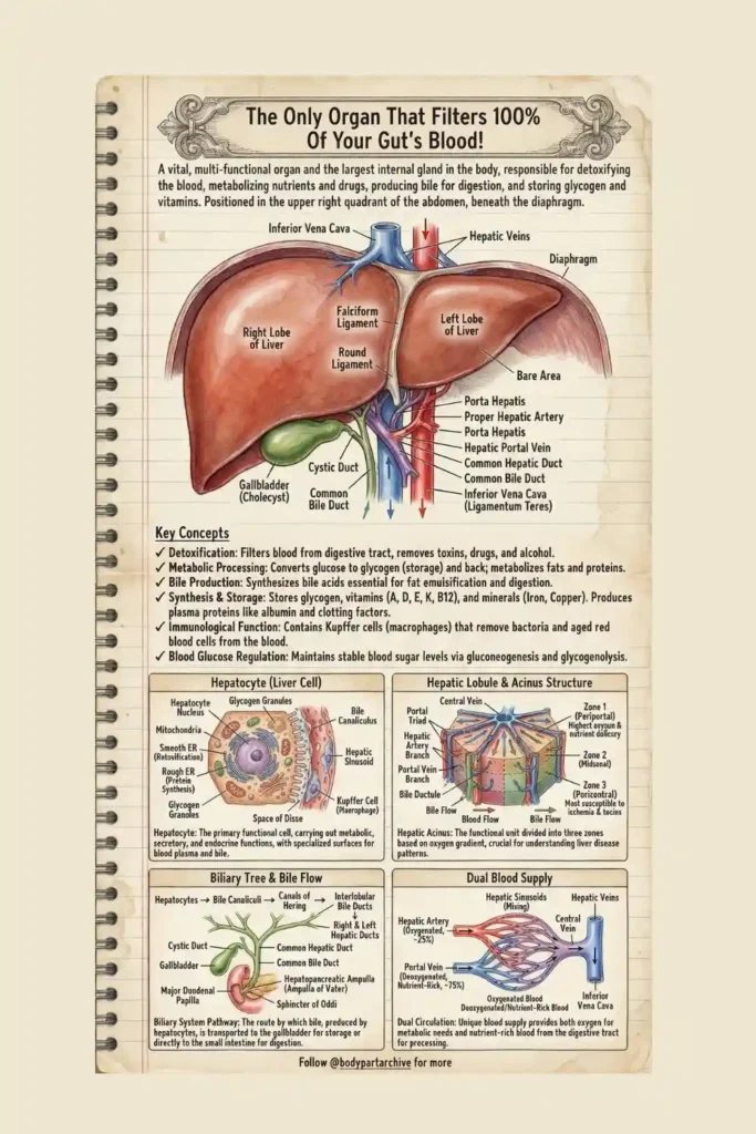
Sitting quietly in the upper right quadrant of your abdomen, safely tucked beneath the diaphragm, is the largest internal gland in the human body: the Liver.
While organs like the heart and lungs have singular, obvious jobs (pumping and breathing), the liver is a master multitasker. It is a highly sophisticated chemical processing plant responsible for over 500 vital functions. It detoxifies the blood, synthesizes proteins, stores essential vitamins, regulates blood sugar, and produces the bile necessary for digestion. Let’s dive into the archives to uncover the microscopic architecture that makes the liver the ultimate biological filter.
The Gross Anatomy & The Biliary Tree
From the outside, the liver is divided into two main visible sections: the large Right Lobe and the smaller Left Lobe, separated by the Falciform Ligament.
Tucked underneath the right lobe is the Gallbladder (Cholecyst), a small, pear-shaped sac. A common misconception is that the gallbladder makes bile. In reality, it is the liver cells that synthesize bile acids (essential for fat emulsification).
This creates a complex plumbing system known as the Biliary Tree. Bile is produced in the hepatocytes, secreted into tiny canals (Bile Canaliculi), and flows down through the Hepatic Ducts. It is then sent to the Gallbladder for storage. When you eat a fatty meal, the bile is released down the Common Bile Duct and through the Sphincter of Oddi into the small intestine to digest your food.
Archive Recommends: Visualizing the Viscera
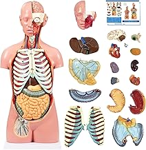
If you are a medical or nursing student struggling to visualize how the liver, gallbladder, and intestines physically connect, textbook 2D images often aren’t enough. We highly recommend using a Amazon Link: 3D Human Torso Anatomy Model on your desk to master the spatial relationships of the abdominal organs.
The Anomaly: A Dual Blood Supply
Most organs have a simple circulatory relationship: an artery brings fresh oxygen, and a vein takes away the waste. The liver, however, has a unique Dual Blood Supply.
The Hepatic Artery: This provides only about 25% of the liver’s blood, bringing the necessary oxygenated blood to keep the liver tissue alive.
The Hepatic Portal Vein: This massive vessel delivers the remaining 75% of the blood. Unlike normal arteries, this blood is deoxygenated. Crucially, it comes directly from the digestive tract! It is extremely nutrient-rich but also packed with raw toxins, alcohol, and absorbed medications.
These two blood supplies physically mix inside the liver’s microscopic channels (Hepatic Sinusoids). This allows the liver to filter and process all the raw materials from your food before that blood is allowed to return to the heart via the Inferior Vena Cava and circulate to your brain.
The Micro-World: Hepatocytes and Kupffer Cells
To perform its massive chemical workload, the liver relies on highly specialized cells.
The primary worker is the Hepatocyte (Liver Cell). Looking closely at a hepatocyte, you will find it packed with organelles. It contains abundant Smooth Endoplasmic Reticulum (for detoxification) and Rough ER (for protein synthesis). You will also see Glycogen Granules. The hepatocyte acts as a battery, converting excess glucose into stored glycogen, and releasing it back into the blood when you are fasting to maintain stable blood sugar levels.
Living inside the sinusoids alongside the hepatocytes are Kupffer Cells. These are specialized macrophages (immune cells). They act as biological security guards, physically engulfing and destroying bacteria and aged red blood cells that pass through the blood.
Archive Recommends: Master Cellular Physiology
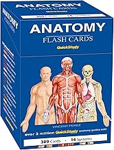
The functions of the smooth ER versus rough ER in hepatocytes are highly tested exam topics. To burn these microscopic details into your memory, utilizing a comprehensive set of Amazon Link: Medical Physiology Flashcards is one of the most effective active-recall strategies you can use.
Clinical Pathology: The Hepatic Acinus & Zone 3
In medical pathology, understanding the liver requires looking at the Hepatic Acinus—the functional unit divided into three zones based on the oxygen gradient.
As mixed blood flows from the portal triad toward the central vein, the oxygen gets depleted:
Zone 1 (Periportal): The cells closest to the incoming blood. They receive the highest levels of oxygen and nutrients.
Zone 2 (Midzonal): The intermediate zone.
Zone 3 (Pericentral): The cells at the very end of the line.
Because Zone 3 receives the least amount of oxygen under normal conditions, it is highly vulnerable. In clinical scenarios, Zone 3 is the most susceptible area to ischemia (lack of oxygen) and damage from toxins (like a Tylenol/acetaminophen overdose). Understanding these zones is critical for diagnosing where and why liver tissue is failing.
Conclusion
The liver is an absolute powerhouse of survival. From manufacturing the bile that digests our food to acting as a 24/7 immune filter and chemical detoxifier, its complex microscopic architecture is beautifully designed to keep the rest of the body safe. Protecting your liver by being mindful of alcohol consumption, medication doses, and maintaining a healthy diet ensures this massive internal filter can continue to perform its 500+ daily miracles.