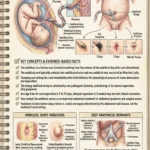
Most people rarely think about their belly button, viewing it as just a quirky feature of their stomach. However, the umbilicus is an anatomical masterpiece. It represents the site of your very first major wound and subsequent scar formation, marking the incredible transition from fetal to independent circulation.
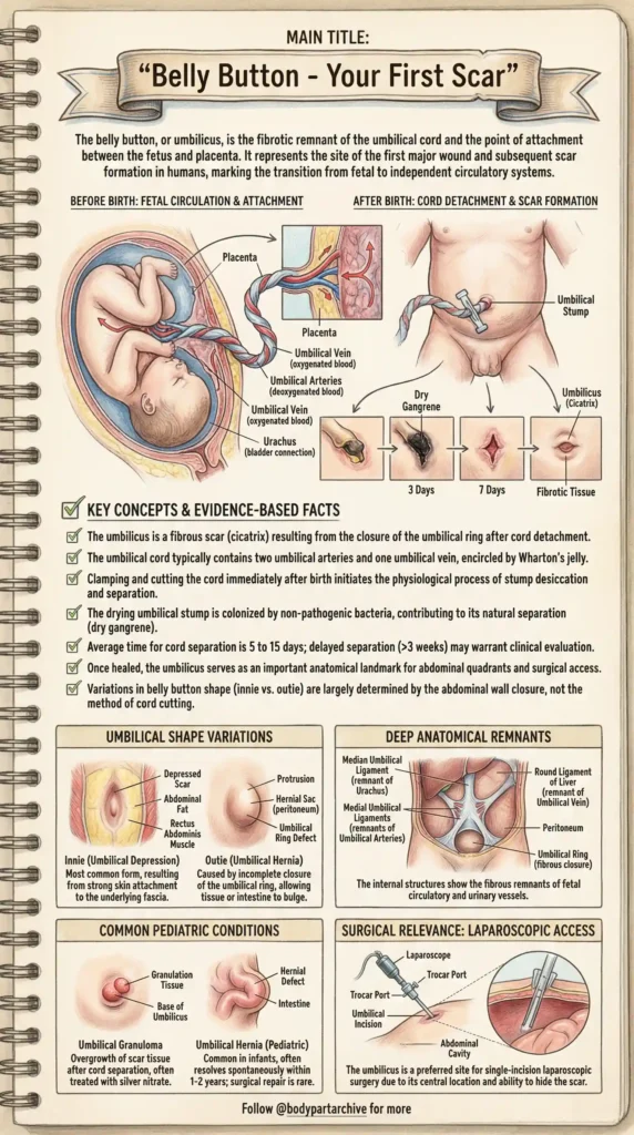
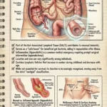
The Origin: Fetal Circulation and Detachment Before birth, the umbilical cord is your lifeline, containing two umbilical arteries (carrying deoxygenated blood) and one umbilical vein (carrying oxygenated blood), all encased in Wharton’s jelly. Clamping and cutting this cord immediately after birth initiates a physiological process. The stump undergoes desiccation and colonization by non-pathogenic bacteria, leading to natural separation (dry gangrene) usually within 5 to 15 days, leaving behind a fibrotic tissue scar.
Innies vs. Outies: What Determines the Shape? Contrary to popular belief, the shape of your belly button has nothing to do with how the doctor cut the cord!
- The “Innie” (Umbilical Depression): This is the most common form, resulting from a strong attachment of the skin to the underlying abdominal fascia.
- The “Outie” (Umbilical Hernia): This is often caused by an incomplete closure of the umbilical ring, allowing a small amount of tissue or intestine to bulge outward.
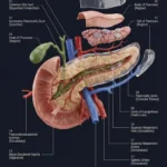
Deep Anatomical Remnants The umbilicus isn’t just a surface scar; it anchors deep internal structures. It hides the fibrous remnants of fetal vessels, such as the Median Umbilical Ligament (remnant of the Urachus) and the Round Ligament of the Liver (remnant of the umbilical vein).
Clinical and Surgical Significance Beyond its fascinating origins, the belly button serves as a crucial anatomical landmark. It divides the abdomen into quadrants and is the preferred site for single-incision laparoscopic surgery, allowing surgeons to access the abdominal cavity while perfectly hiding the surgical scar.