The Pelvis Structure: Exploring The Body’s Foundation
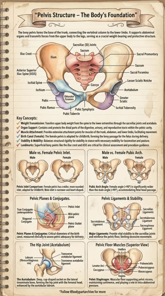
The bony pelvis acts as the central architectural foundation of the human body. Serving as a massive, sturdy ring of bone, it forms the base of the trunk, connecting the vertebral column to the lower limbs. For students of medicine, nursing, physical therapy, and obstetrics, understanding the complex topography of the pelvis is absolutely essential. The pelvis does not merely sit passively; it is a highly dynamic structure responsible for weight transmission, protecting delicate internal organs (digestive, urinary, and reproductive tracts), and providing extensive attachment points for the powerful muscles of the trunk and legs. Today, we are breaking down the anatomy of the pelvis using our beautifully illustrated, vintage-style study guide.
The Bony Landmarks: Ilium, Ischium, and Pubis
During childhood, each side of the pelvis consists of three separate bones: the Ilium, the Ischium, and the Pubis. By adulthood, these bones fuse completely to form a single, solid “innominate” (hip) bone. These two hip bones meet anteriorly at the pubic symphysis and attach posteriorly to the sacrum at the sacroiliac (SI) joints.
- The Ilium: The largest and most superior part of the pelvis. Its flared upper border is the Iliac Crest, a major superficial landmark you can feel on your waist. The front edge features the Anterior Superior Iliac Spine (ASIS), a crucial reference point for clinical assessments and surgical procedures.
- The Ischium: The posterior and inferior portion of the pelvis. It features the prominent Ischial Tuberosity—often referred to as your “sit bone” because it bears your weight when you sit down.
- The Pubis: The anterior portion of the pelvis. The left and right pubic bones meet in the midline at a cartilaginous joint called the Pubic Symphysis.
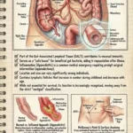
Male vs. Female Pelvic Differences
One of the most highly tested subjects in anatomy is the sexual dimorphism of the human pelvis. Because the female pelvis must accommodate the passage of a fetal head during childbirth, it is structurally distinct from the male pelvis. The Pelvic Inlet: * Female: The inlet is wider, more spacious, and generally rounded or oval-shaped to allow for fetal descent.
- Male: The inlet is narrower, more robust, and characteristically heart-shaped due to the prominent sacral promontory projecting into it.
The Pubic Arch:
- Female: The angle formed below the pubic symphysis is broad and wide, measuring significantly greater than 90 degrees.
- Male: The angle is much sharper and narrower, typically measuring less than 90 degrees.
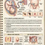
Pelvic Planes, Conjugates, and Obstetrics
For obstetrics and midwifery, simply knowing the bones is not enough; one must understand the internal dimensions of the birth canal. Clinical measurements called “conjugates” are used to assess whether a pelvis is adequate for vaginal delivery.
- Pelvic Inlet & Outlet: These imaginary planes define the upper and lower boundaries of the true pelvic cavity.
- True Conjugate (Anteroposterior): The distance from the top of the pubic symphysis to the sacral promontory.
- Diagonal Conjugate: Measured clinically during a pelvic exam from the inferior margin of the pubic symphysis to the sacral promontory. It helps physicians estimate the true conjugate.
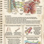
Ligamentous Stability and The Acetabulum
The pelvis must withstand massive forces, especially during walking or running. This stability is reinforced by some of the strongest ligaments in the human body.
- Major Ligaments: The Sacroiliac ligaments lock the sacrum to the ilium. Deeper down, the Sacrotuberous and Sacrospinous ligaments prevent the sacrum from tilting forward under the weight of the spine, essentially locking the pelvic ring in place.
- The Hip Joint (Acetabulum): Where the three pelvic bones (Ilium, Ischium, Pubis) fuse, they form a deep, cup-shaped socket called the acetabulum. The head of the femur fits tightly into this socket, further deepened by a ring of fibrocartilage called the acetabular labrum, creating a highly stable and mobile hip joint.
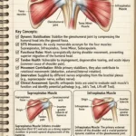
The Hidden Support: Pelvic Floor Muscles
Looking at the pelvis from a superior view, you can see it is not empty; its base is sealed by a muscular sling known as the pelvic diaphragm.
The Levator Ani Group: This powerful muscle group consists of the Puborectalis, Pubococcygeus, and Iliococcygeus. Together with the smaller Coccygeus muscle, they form a supportive hammock.
- Function: The pelvic floor muscles support the pelvic viscera (bladder, intestines, uterus), maintain urinary and fecal continence, and play a vital role in managing intra-abdominal pressure during heavy lifting or coughing.
Conclusion
The pelvis is a structural masterpiece, perfectly balancing the need for rigid stability with necessary mobility. By breaking down its complex anatomy visually—from the superficial bony landmarks to the deep pelvic floor muscles—students can master this high-yield topic with much less stress.
Bookmark this vintage anatomical guide for your next anatomy exam, and be sure to explore BodyPartArchive for more visual cheat sheets that make medical science beautiful and easy to understand!