Introduction: The Organ with an Identity Crisis
Tucked away deep in the upper abdomen, sitting horizontally behind the stomach, lies one of the most remarkable and complex organs in the human body: the pancreas. For medical, nursing, and biology students, the pancreas often presents a unique studying challenge because it is essentially an organ with an identity crisis. It performs two entirely different, full-time jobs.
It acts as an exocrine gland to facilitate the digestion of the food we eat, and simultaneously functions as an endocrine gland to regulate our blood sugar levels. Attempting to understand how these two disparate systems operate within the same physical space using flat, 2D textbook diagrams can be incredibly confusing. By utilizing a high-yield, exploded 3D visual master diagram, we can peel back the layers of the pancreas to understand its regional anatomy, functional tissues, and crucial vascular supply.
Regional Anatomy: From Head to Tail
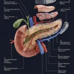
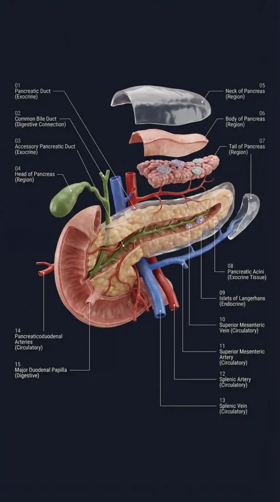
Before diving into the microscopic functions, it is essential to understand the gross regional anatomy of the pancreas. As highlighted in our 3D visualization, the pancreas is divided into four main sections:
- Head of Pancreas (04): The widest part, nestled snugly into the C-shaped curve of the duodenum (the first part of the small intestine).
- Neck of Pancreas (05): A short, constricted region connecting the head to the body.
- Body of Pancreas (06): The main, elongated central portion that stretches behind the stomach.
- Tail of Pancreas (07): The narrow, tapered end that points towards the spleen on the left side of the abdomen.
Understanding these regions is clinically significant; for example, pancreatic cancer most frequently originates in the head of the pancreas, which can quickly compress adjacent structures like the bile duct.
The Digestive Factory: The Exocrine Pancreas
Approximately 95% of the pancreas is dedicated to its exocrine function—digestion.
Look closely at the yellow, lobulated tissue in the 3D diagram labeled Pancreatic Acini (08). These acinar cells are the tiny biological factories that produce powerful digestive enzymes (amylase, lipase, and proteases). Once produced, these enzymes are secreted into a network of tiny tubes that eventually merge into the main Pancreatic Duct (01).
Our diagram brilliantly illustrates how this duct runs down the entire length of the organ. At the head of the pancreas, it is joined by the green Common Bile Duct (02), which brings bile down from the liver and gallbladder. Together, these two critical fluids exit into the duodenum through a specialized opening called the Major Duodenal Papilla (15). This shared doorway is a high-yield clinical concept: if a migrating gallstone becomes lodged at this papilla, it blocks the flow of both bile and pancreatic juice. The trapped pancreatic enzymes can become activated prematurely, beginning to digest the pancreas itself—a painful and dangerous condition known as acute gallstone pancreatitis.
The Blood Sugar Control Room: The Endocrine Pancreas
Embedded within the vast sea of exocrine acinar cells are tiny, highly vascularized clusters of cells. In our 3D model, these are highlighted as the purple cellular islands labeled the Islets of Langerhans (09).
Despite making up only 1% to 2% of the pancreatic mass, these islets are responsible for the organ’s critical endocrine function. They contain different types of cells, most notably:
- Alpha cells: Produce glucagon, which raises blood sugar levels when they drop too low.
- Beta cells: Produce insulin, which lowers blood sugar levels by allowing cells to absorb glucose from the bloodstream.
Unlike the exocrine system, which uses ducts, the endocrine system is ductless. The Islets of Langerhans secrete insulin and glucagon directly into the surrounding blood vessels to be distributed throughout the entire body. When these beta cells are destroyed (Type 1 Diabetes) or when the body becomes resistant to the insulin they produce (Type 2 Diabetes), the body loses its ability to regulate blood glucose.
The Vascular Network: Feeding the Pancreas
Because of its intense endocrine and exocrine workload, the pancreas requires a robust blood supply. Our exploded 3D view meticulously maps out this complex vasculature.
The Splenic Artery (12), branching off from the celiac trunk, follows a tortuous path along the upper border of the pancreas, supplying the body and tail before reaching the spleen. The corresponding Splenic Vein (13) runs right behind the pancreas. Meanwhile, the lower portions and head are supplied by the Pancreaticoduodenal Arteries (14) and closely associated with the massive Superior Mesenteric Artery (11) and Superior Mesenteric Vein (10). Visualizing how these major vessels intertwine with the pancreatic tissue is crucial for medical students, especially those entering surgical rotations where protecting these vessels during pancreatic or abdominal surgery is paramount.
Conclusion: Visualizing Complexity
The pancreas is a masterclass in biological efficiency, housing both a digestive enzyme factory and a hormonal control center within a single, elegant structure. Staring at dense paragraphs of text to memorize the relationship between the Acini, the Islets, and the biliary tree is an outdated study method.
(Are you struggling to connect gross anatomy with complex physiological functions? The detailed “3D Exploded Pancreas” master diagram featured in this article is just ONE page from our massive Complete 50+ Visual Anatomy Notes Bundle. We have meticulously mapped out the entire human body—covering Gastrointestinal, Endocrine, Neuroanatomy, and Cardiovascular Systems—into beautiful, highly structured visual study guides. Stop reading walls of text and start learning visually. Visit our digital archive today to download the ultimate Visual Study Kit and upgrade your medical education!)