Disclosure: As an Amazon Associate, Body Part Archive earns from qualifying purchases. This means if you click on a link and buy something, we may earn a small commission at no extra cost to you.
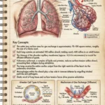
Introduction: The Tennis Court in Your ChestBreathing is so automatic that we rarely stop to appreciate the mechanical and structural miracles happening inside our chests roughly 20,000 times a day. The human lungs are not just hollow bags that fill with air; they are incredibly intricate, highly vascularized biological sponges designed for one critical mission: to swap out toxic carbon dioxide for life-sustaining oxygen.
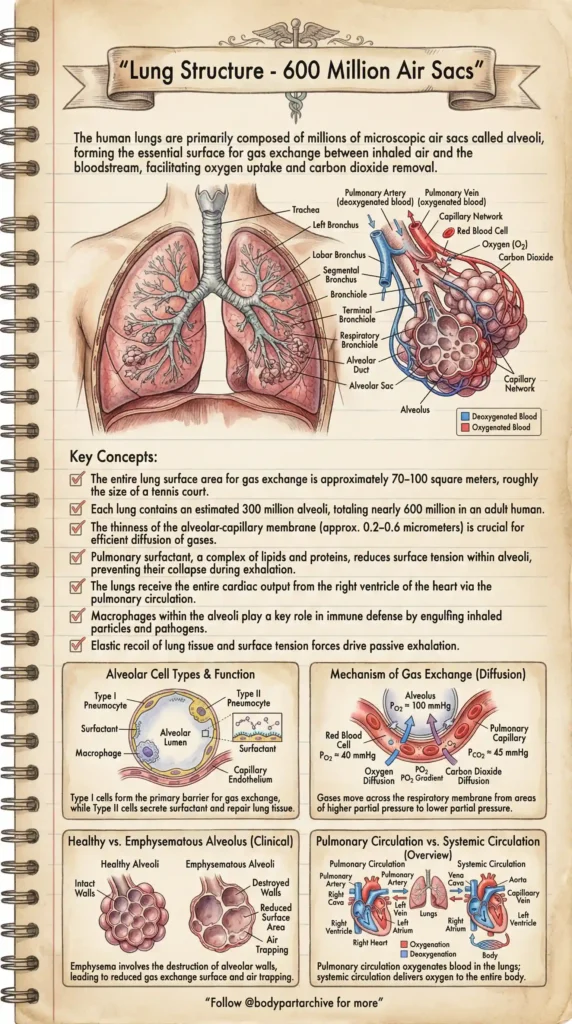
To achieve this, the lungs rely on microscopic structures called Alveoli. You have an estimated 300 million of these air sacs in each lung, totaling nearly 600 million. If you were to flatten out the incredibly thin walls of every alveolus, the total surface area for gas exchange would span 70 to 100 square meters—roughly the size of a tennis court! Let’s dive into the archives to explore the anatomy, physics, and pathology of the respiratory system.
The Respiratory Tree: From Trachea to Alveoli
When you inhale, air travels down the Trachea (windpipe), which splits into the left and right Bronchi. These tubes continue to branch into smaller and smaller pathways—Lobar Bronchi, Segmental Bronchi, and finally, tiny Bronchioles.
At the very end of the Terminal Bronchioles, the airway opens into Alveolar Ducts and clusters of Alveolar Sacs. This is where the magic happens. The entire lung receives the total cardiac output from the right ventricle of your heart, meaning every drop of your blood is pumped through the capillary networks wrapping these sacs.
Archive Recommends: Lung Health & Capacity
To keep your alveoli actively expanding and clear of mucus, respiratory therapists often recommend deep breathing exercises. A simple [ Volumetric Incentive Spirometer] is an excellent, inexpensive tool to train your lung capacity and keep your respiratory muscles strong.Archive Recommends: Lung Health & Capacity
The Microscopic Miracle: Alveolar Cell Types
Zooming in on a single alveolus reveals a highly specialized microscopic environment. The walls of these sacs are composed of distinct cell types:
Type I Pneumocytes: These form the primary barrier for gas exchange. They are incredibly thin, allowing the alveolar-capillary membrane to be just 0.2-0.6 micrometers thick, which is essential for rapid gas diffusion.
Type II Pneumocytes: These are the unsung heroes of the lung. They secrete a crucial substance called Pulmonary Surfactant (a complex of lipids and proteins). Surfactant coats the inside of the alveolus to reduce surface tension. Without it, the wet walls of the air sacs would stick together, and your lungs would collapse during exhalation.
Macrophages: The biological security guards. These immune cells patrol the alveolar lumen, engulfing and destroying inhaled dust particles and pathogens that managed to bypass the upper airways.
The Physics of Breathing: Mechanism of Gas Exchange
How does oxygen actually leap from the air into your blood? It all comes down to Diffusion and partial pressure gradients. Gases naturally move from areas of higher pressure to areas of lower pressure.
When fresh air reaches the alveolus, the partial pressure of oxygen (PO2) is high (about 100 mmHg). Meanwhile, the deoxygenated blood arriving in the pulmonary capillaries from the body has a much lower PO2 (about 40 mmHg). Because of this steep gradient, oxygen diffuses instantly across the membrane into the red blood cells. Conversely, Carbon Dioxide (PCO2) is higher in the blood than in the fresh air, so it diffuses out into the alveolus to be exhaled.
Clinical Pathology: The Devastation of Emphysem
While the lungs are resilient, their delicate microscopic architecture is highly vulnerable to inhaled toxins, particularly cigarette smoke.
A severe consequence of chronic smoking is Emphysema. Looking at a clinical comparison, healthy alveoli have intact walls, maintaining that massive “tennis court” surface area. In emphysema, the toxic insult causes the destruction of these walls. The clusters of tiny “grapes” merge into large, floppy, inefficient spaces.
This destruction has two devastating effects:
Reduced Surface Area: Less membrane means significantly less oxygen can diffuse into the bloodstream.
Air Trapping: Healthy lungs rely on the “elastic recoil” of tissue to push air out passively. Emphysema destroys this elasticity. The airways collapse during exhalation, trapping stale air inside the lungs and making the simple act of breathing out incredibly laborious.
Conclusion
Your 600 million alveoli are the fragile, microscopic engine of your life force. From the precise secretion of surfactant to the physics of gas diffusion, the respiratory system is a perfectly balanced machine. Protecting this system—by avoiding smoking, minimizing exposure to pollutants, and engaging in cardiovascular exercise—is paramount to maintaining that massive, life-giving surface area for decades to come.
Disclosure: As an Amazon Associate, Body Part Archive earns from qualifying purchases. This means if you click on a link and buy something, we may earn a small commission at no extra cost to you.