Introduction: The Ultimate Biological Highway
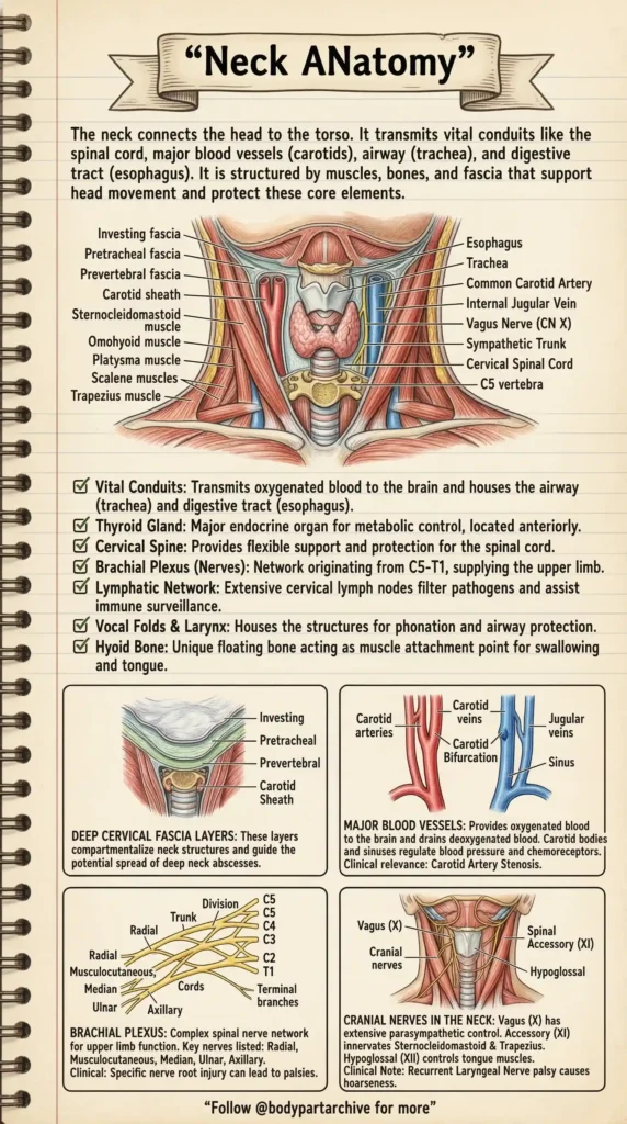
The human neck is arguably the most complex and crowded region of the entire body. It serves as the vital bridge connecting the command center (the head) to the rest of the torso. Despite its relatively small size, it must safely transmit the spinal cord, major blood vessels like the carotids, the airway (trachea), and the digestive tract (esophagus).
Disclosure: As an Amazon Associate, Body Part Archive earns from qualifying purchases. This means if you click on a link and buy something, we may earn a small commission at no extra cost to you.
Because it lacks the bony protection of a ribcage, the neck relies on a highly engineered system of muscles, cervical bones, and tough fascia to support head movement and protect these core elements. Let us dive into the clinical archives and unpack the brilliant anatomy of the neck.
The Vital Conduits and Structures
Looking at an anterior cross-section of the neck reveals a masterclass in spatial efficiency. Hidden beneath major muscles like the Sternocleidomastoid and the Trapezius are several critical structures:
The Thyroid Gland: A major endocrine organ located anteriorly, responsible for metabolic control.
The Airway and Digestive Tract: The trachea (windpipe) and esophagus run directly down the centerline. Sitting just above them are the Vocal Folds and Larynx, which house the structures required for phonation (speaking) and airway protection.
The Hyoid Bone: A fascinating anatomical anomaly. The hyoid is a unique “floating” bone. It acts as a crucial muscle attachment point for swallowing and supporting the tongue.
Archive Recommends: Protect Your Cervical Spine
The neck is highly susceptible to postural strain (like “Tech Neck”). To maintain the natural curve of your cervical spine and relieve tension on the Sternocleidomastoid and Trapezius muscles, we highly recommend switching to an [Amazon Link: Ergonomic Cervical Memory Foam Pillow] for proper nighttime support.
The Architecture: Deep Cervical Fascia Layers
The neck is not a hollow tube; it is strictly organized into compartments by tough layers of connective tissue known as the Deep Cervical Fascia.
These fascial layers include the Investing, Pretracheal, and Prevertebral fascia, as well as the Carotid Sheath. Clinically, these layers are of paramount importance. They compartmentalize the neck structures. If a patient develops a severe infection (like a deep neck abscess), these fascial planes will guide the potential spread of the infection, sometimes directing it downwards into the highly dangerous thoracic (chest) cavity.
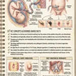
Blood Supply: The Carotid and Jugular Networks
Your brain consumes a massive amount of energy, and the neck is responsible for delivering its fuel. The Major Blood Vessels of the neck include the Carotid arteries (delivering oxygenated blood to the brain) and the Jugular veins (draining deoxygenated blood).
At the Carotid Bifurcation (where the artery splits), there are specialized structures called Carotid bodies and sinuses. These act as biological sensors to regulate blood pressure and function as chemoreceptors. Clinically, conditions like Carotid Artery Stenosis (narrowing of these arteries) can severely impact blood flow and lead to strokes.
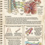
The Wiring: Cranial Nerves and the Brachial Plexus
Finally, the neck is a major neurological transit zone.
Cranial Nerves:
Several cranial nerves travel through the neck, including the Vagus Nerve (CN X), which provides extensive parasympathetic control to the body’s internal organs. The Accessory Nerve (CN XI) innervates the Sternocleidomastoid and Trapezius muscles, while the Hypoglossal Nerve (CN XII) controls the muscles of the tongue. A clinical note to remember: palsy of the Recurrent Laryngeal Nerve (a branch of the Vagus) causes hoarseness of the voice.
The Brachial Plexus:
Perhaps most importantly for upper body mobility, the neck houses the origin of the Brachial Plexus. This complex network originates from the C5 to T1 spinal nerve roots. It divides into trunks, divisions, and cords before forming the terminal branches (Radial, Musculocutaneous, Median, Ulnar, and Axillary nerves) that supply the entire upper limb. This is why a specific nerve root injury in the neck can lead to severe palsies, pain, or numbness in the hand or arm.
Archive Recommends: Master the Nerves
The Brachial Plexus is notoriously difficult to memorize for medical exams. We strongly suggest grabbing a set of [Amazon Link: Netter’s Anatomy Flashcards] to help visualize and memorize these complex nerve pathways and clinical correlations.
Conclusion
From the protective layers of deep fascia to the incredible wiring of the brachial plexus, the neck is a structural marvel. Understanding its anatomy is not just for passing medical exams; it is essential for diagnosing pain, understanding the spread of infections, and appreciating how a single compromised nerve in the cervical spine can affect the entire upper limb.